

Why get dental X-rays before major treatments
When it comes to major dental treatment, hidden details can make all the difference. From the shape of your tooth roots to the condition of your jawbone, what lies beneath the surface can influence how treatment is planned and performed.
Dental X-rays allow your dentist to see these hidden structures, revealing potential problems and important anatomical details that cannot be assessed by sight alone.
This becomes particularly important before major treatments such as dental implants, orthodontics, root canals and complex extractions. Imaging provides valuable information that can help your dentist understand your oral health and plan treatment appropriately.
What lies beneath
A visual examination allows your dentist to assess the visible surfaces of your teeth, gums and other tissues. It can reveal signs of decay, inflammation, damage and changes to your bite.
Some problems, however, develop beneath the surface.
X-rays can show tooth roots, supporting bone, developing or impacted teeth and changes around the tips of roots. They can also help identify cavities between teeth that may be difficult to detect visually and provide information about areas where infection or bone loss may be present.
Importantly, dental problems do not always cause pain. An issue can develop quietly and only become apparent when it has progressed. Identifying it before major treatment gives your dentist an opportunity to consider it as part of your overall treatment plan.
How X-rays guide major treatment
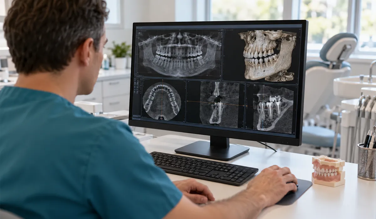
From moving teeth into alignment to placing implants or performing delicate extractions, every dental procedure presents its own challenges. X-rays help dentists understand the anatomy involved, identify potential complications and plan an appropriate approach.
Orthodontics
Braces and clear aligners move teeth through the supporting bone, so understanding the position of the teeth and their roots is an important part of treatment planning.
X-rays can help assess root development, the position of unerupted or impacted teeth and the surrounding bone. For more complex orthodontic treatment, additional imaging may be considered to provide information about the jaws and other structures.
This can be particularly relevant when treatment involves significant tooth movement or appliances designed to influence the width or position of the upper jaw.
Dental implants
Dental implants are placed into the jawbone, making the condition and anatomy of the proposed implant site important considerations.
Imaging can help your dentist assess the available bone and the location of nearby structures, such as nerves or the maxillary sinus. In more complex cases, a three-dimensional CBCT scan may provide additional information for treatment planning.
The aim is to understand the implant site and surrounding anatomy before treatment begins, rather than relying only on what can be seen clinically.
Root canals
Root canal treatment involves cleaning and sealing the canals inside a tooth. These canals can vary in number, shape and direction from one tooth to another.
An X-ray can help the dentist assess the roots and surrounding bone and identify changes that may indicate infection or inflammation.
Imaging may also be used during and after treatment to assist with planning and assessment.
Tooth extractions
The visible part of a tooth does not always tell the whole story. Roots can be curved, unusually shaped or positioned close to important structures.
An X-ray can help your dentist understand the shape and position of the roots before an extraction. This is particularly relevant for wisdom teeth, which may be positioned close to nerves or other anatomical structures.
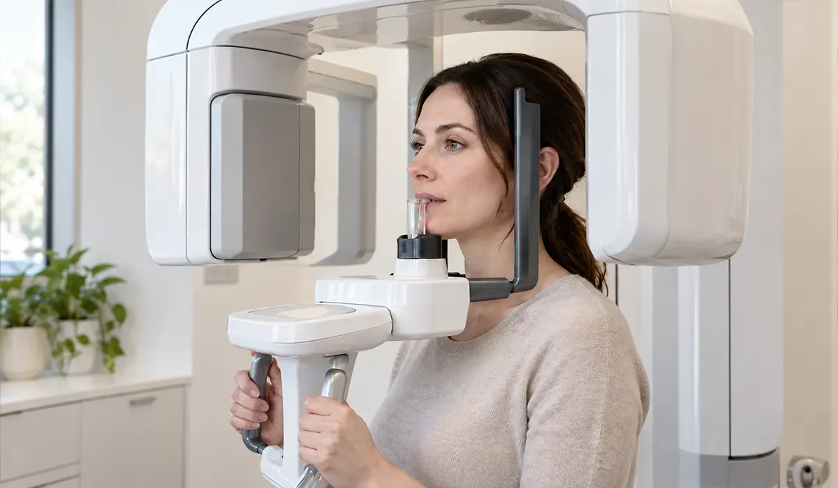
Which type of dental X-ray do you need?
Dental X-rays serve different purposes, from detecting decay between teeth to providing detailed three-dimensional views of the jaw. Your dentist will recommend the most appropriate imaging based on what needs to be investigated and the treatment being considered.
Bitewing X-rays provide detailed images of the crowns of the upper and lower teeth. They are commonly used to detect decay between teeth and assess supporting bone levels.
Periapical X-rays show an entire tooth, including its root and surrounding bone. They can be useful when investigating pain, infection, trauma or changes around a tooth’s root.
Panoramic X-rays provide a broad view of the upper and lower jaws in one image. They can help assess the position and development of teeth, including unerupted or impacted teeth.
CBCT scans create three-dimensional images of the teeth, jawbone and surrounding structures. They may be recommended when more detailed information is needed for complex treatment planning, such as certain implant or surgical procedures.
More imaging is not necessarily better. Your dentist will recommend the type of imaging that provides useful information for your particular situation.
Are dental X-rays safe?
Modern dental X-rays offer an impressive balance of diagnostic detail and low radiation exposure. While it’s natural to have questions about safety, the doses involved are generally very small compared with everyday sources of radiation.
To put this into perspective, some dental X-rays involve less radiation than the natural background radiation we receive from our surroundings in a single day.
Even with these low exposure levels, your dental team takes every precaution to keep radiation exposure to a minimum. X-rays are recommended only when clinically justified, using the lowest practical dose needed to obtain clear, useful images. This follows a principle known as ALARA, meaning “as low as reasonably achievable.”
Modern digital X-rays can also reduce radiation exposure compared with traditional film, while producing images that your dentist can review almost immediately.
Your dentist may also check your previous X-rays before recommending new ones. If existing images still provide the information needed, there may be no reason to repeat them.
If you’re pregnant, think you might be pregnant or simply feel uneasy about having an X-ray, don’t hesitate to speak with your dental team. They’ll be happy to explain the benefits, discuss any concerns and help you understand what’s appropriate for your individual circumstances.
The bigger picture of your oral health

Dental X-rays are just one part of understanding your oral health. Your dentist will also consider your dental history, symptoms and clinical findings, along with any photographs, scans or other information relevant to your care.
Together, these details help build a more complete picture of your teeth, gums and surrounding structures, allowing your dentist to make informed recommendations tailored to your needs.
X-rays can also help you better understand your own oral health. By showing you what’s happening beneath the surface, your dentist can explain their findings, discuss your treatment options and help you feel more confident about the decisions ahead.
Better information for better planning
The fewer surprises during dental treatment, the better. While no procedure is entirely predictable, careful preparation can help your dentist anticipate challenges and make informed decisions before treatment begins.
Dental X-rays play an important role in this preparation, revealing hidden problems and providing valuable information about your tooth roots, jawbone and surrounding structures. They can also help identify issues that may need attention beforehand, allowing your dentist to approach complex procedures with a clearer understanding of what to expect.
Protect your smile from the inside out
Looking after your teeth is not only about treating problems when they appear. Regular dental examinations, appropriate diagnostic imaging and consistent oral hygiene all have a role in maintaining long-term oral health.
Brushing twice a day with fluoride toothpaste, cleaning between your teeth and attending regular dental appointments can help prevent or detect problems before they become more complex.
If you are considering major dental treatment, speak with your dentist about the examinations and imaging that may be appropriate for your individual needs. Understanding what is happening beneath the surface can be an important part of making informed decisions and maintaining a healthy smile for years to come.